Endometrial Cancer
- Home
- »
- Cancer Info
- »
- Endometrial Cancer

Cancer Treatment
Endometrial Cancer
Symptoms of Endometrial Cancer:
The most common symptom of endometrial cancer is abnormal vaginal bleeding. It can be
- Abdominal lumps
- Abdominal pain and bloating
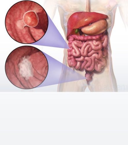
- Any changes in bowel habits
- Blood from the rectum
- Blood in faces, making it look black
- Constipation
- Diarrhoea
- Fatigue
- A feeling of improper bowel movements
- Iron deficiency
- Loss of appetite
- Unexplained weight loss

Cancer Treatment
Types of Endometrial Cancer
There are two types of endometrial carcinoma:
- Type I or endometroid adenocarcinoma is associated with excess estrogen secretion less aggressive type and spread relatively slowly.
- Type II or serous carcinoma or clear cell carcinoma are non-estrogen dependant and aggressive types of cancer.

Cancer Treatment
Causes of Endometrial Cancer:
Endometrium growth varies according to the levels of estrogen and progesterone hormones secreted from ovaries. Excess levels of estrogen are responsible for the proliferation of tumours in the endometrium, causing Type I and Type II endometrial cancers.
Non-modifiable
- Age: Chances of contracting cancer increase as we age.
- Family history: About 5-10% of endometrium cancers are related to family history. History of multiple family members having colon cancer increases the possibility of familial inheritance and should be ruled out with genetic testing; Lynch syndrome is a condition where the chances of multiple family members having various cancers are high.
- Early menarche and late menopause increase the duration of menstruation in years, exposing the endometrium to the estrogen hormone for a longer period, which increases the chances of endometrial cancer.
- Estrogen-secreting ovarian cancers can also cause endometrial cancer.
Modifiable Causes
Most of these causes are related to the secretion of excess Estrogen due to external causes.
- Obesity: Once women pass the menstruation phase the ovarian function stops but estrogen hormone can be produced from fat cells known as adipose tissue of the body. This is one of the most common causes of endometrium cancer. Obesity, diabetes and high blood pressure usually coexist in patients and are known as the triple syndrome of endometrial cancer and are responsible for excessive estrogen production in the body.
- Diet and physical activity: High-fat content in diets and sedentary lifestyles are related to increased body weight and in turn are related to endometrial cancer.
- Polycystic ovarian syndrome: This condition is associated with the secretion of excess estrogen and androgen hormones causing endometrial cancer.
- Hormone therapy for breast cancer: Tamoxifen is an anti-cancer drug used for the treatment of breast cancer and can cause endometrial hyperplasia (increase in thickness of endometrial lining). The association of tamoxifen with endometrial cancer has been proven since the chances are not very high and considering the risk-benefit ratio tamoxifen is considered an essential medicine for breast cancer hence the use of same should not be stopped.
Prevention of Endometrial Cancer
- Healthy lifestyle choices
- Weight reduction
- Periodical health check-ups

Investigations to diagnose Endometrial Cancer

Treatment for Endometrial Cancer:
For older women who have had children past their childbearing age the following treatment options are available:
Surgery:
Radiation therapy:
- Based on histopathology findings, the need for radiation therapy is determined, and early-stage cancers do not require further treatment. Brachytherapy is an internal radiation therapy used for cancers spread deep inside the uterus.
- External beam radiation therapy (EBRT) is used to treat tumours, which has spread to the lymph nodes.
Chemotherapy:
The need for chemotherapy is determined based on histopathological findings and is given to patients to avoid further spread of tumours.
Hormone therapy:
The need for chemotherapy is determined based on histopathological findings and is given to patients to avoid further spread of tumours.
Testimonial
Feedback From Our Happy Patients


